
Page 51 - IMCJ19s1
P. 51
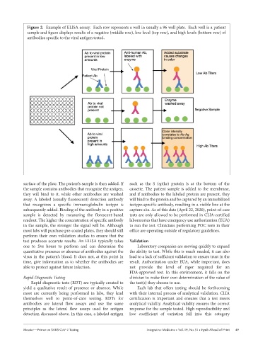
Figure 2.  Example of ELISA asssay.  Each row represents a well in usually a 96 well plate.  Each well is a patient
              sample and figure displays results of a negative (middle row), low level (top row), and high levels (bottom row) of
              antibodies specific to the viral antigen tested.


            surface of the plate. The patient’s sample is then added. If   such  as  the  S  (spike)  protein  is  at  the  bottom  of  the
            the sample contains antibodies that recognize the antigen,   cassette. The patient sample is added to the membrane,
            they  will  bind  to  it,  while  other  antibodies  are  washed   and if antibodies to the labeled protein are present, they
            away. A labeled (usually fluorescent) detection antibody   will bind to the protein and be captured by an immobilized
            that  recognizes  a  specific  immunoglobulin  isotype  is   isotype-specific antibody, resulting in a visible line at the
            subsequently added. Binding of the antibody in a positive   capture site. As of this date (April 22, 2020), point-of-care
            sample  is  detected  by  measuring  the  florescent-based   tests are only allowed to be performed in CLIA-certified
            readout. The higher the concentration of specific antibody   laboratories that have emergency use authorization (EUA)
            in the sample, the stronger the signal will be. Although   to run the test. Clinicians performing POC tests in their
            most labs will purchase pre-coated plates, they should still   office are operating outside of regulatory guidelines.
            perform  their  own  validation  studies  to  ensure  that  the
            test  produces  accurate  results.  An  ELISA  typically  takes   Validation
            one  to  five  hours  to  perform  and  can  determine  the   Laboratory companies are moving quickly to expand
            quantitative presence or absence of antibodies against the   the ability to test. While this is much needed, it can also
            virus in the patient’s blood. It does not, at this point in   lead to a lack of sufficient validation to ensure trust in the
            time,  give  information  as  to  whether  the  antibodies  are   result. Authorization under EUA, while important, does
            able to protect against future infection.        not  provide  the  level  of  rigor  required  for  an
                                                             FDA-approved  test.  In  this  environment,  it  falls  on  the
            Rapid Diagnostic Testing                         clinician to make their own determination of the value of
               Rapid diagnostic tests (RDT) are typically created to   the test(s) they choose to use.
            yield  a  qualitative  result  of  presence  or  absence.  While   Each  lab  that  offers  testing  should  be  forthcoming
            most  are  currently  being  performed  in  labs,  they  lend   with their internal process of analytical validation. CLIA
            themselves  well  to  point-of-care  testing.  RDTs  for   certification  is  important  and  ensures  that  a  test  meets
            antibodies  are  lateral  flow  assays  and  use  the  same   analytical validity. Analytical validity ensures the correct
            principles  as  the  lateral  flow  assays  used  for  antigen   response for the sample tested. High reproducibility and
            detection discussed above. In this case, a labeled antigen   low  coefficient  of  variation  fall  into  this  category


            Messier—Primer on SARS-CoV-2 Testing                     Integrative Medicine • Vol. 19, No. S1 • Epub Ahead of Print  49