
Page 52 - IMCJ19s1
P. 52
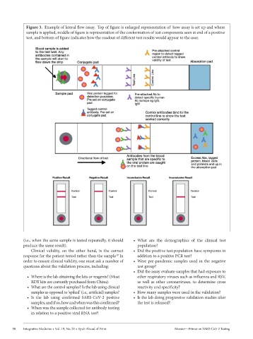
Figure 3.  Example of lateral flow assay.  Top of figure is enlarged representation of  how assay is set up and where
             sample is applied, middle of figure is representation of the conformation of test components seen at end of a positive
             test, and bottom of figure indicates how the readout of different test results would appear to the user.


            (i.e., when the same sample is tested repeatedly, it should   •  What  are  the  demographics  of  the  clinical  test
            produce the same result).                            population?
               Clinical  validity,  on  the  other  hand,  is  the  correct   •  Did the positive test population have symptoms in
            response for the patient tested rather than the sample  In   addition to a positive PCR test?
                                                       19
            order to ensure clinical validity, one must ask a number of   •  Were  pre-pandemic  samples  used  in  the  negative
            questions about the validation process, including:   test group?
                                                               •  Did the assay evaluate samples that had exposure to
              •  Where is the lab obtaining the kits or reagents? (Most   other respiratory viruses such as influenza and RSV,
               RDT kits are currently purchased from China).     as  well  as  other  coronaviruses,  to  determine  cross
              •  What are the control samples? Is the lab using clinical   reactivity and specificity?
               samples as opposed to ‘spiked’ (i.e., artificial) samples?  •  How many samples were used in the validation?
              •  Is  the  lab  using  confirmed  SARS-CoV-2  positive   •  Is the lab doing progressive validation studies after
               samples, and if so, how and when was this confirmed?  the test is released?
              •  When was the sample collected for antibody testing
               in relation to a positive viral RNA test?


       50   Integrative Medicine • Vol. 19, No. S1 • Epub Ahead of Print          Messier—Primer on SARS-CoV-2 Testing